Keratoconus: are the first warning signs in our genes?

csm-0719-700-webfirst-avellino-in-article-1599808f09_orig
A new tool will soon be available to ophthalmologists, it will use NGS sequencing technology and a custom genetic panel for early detection of the disease.

Eye doctors dealing with keratoconus are about to benefit from a new tool at their disposal; Avellino Labs is developing a diagnostic genetic test for risk factors for keratoconus.

The new technology of Avellino Labs It will not only offer ophthalmologists a tool for early diagnosis of patients at risk of developing keratoconus but will also provide additional data for patients who may not show the classic signs associated with keratoconus when examined using current scanning technologies and algorithms.
Edward J. Holland, Director of Cornea Services at the Cincinnati Eye Institute and Professor of Ophthalmology at the University of Cincinnati, says, “Adding genetic testing to our existing optical and scanning methods will result in the ability to identify patients at risk for keratoconus earlier, perhaps before there are changes that can be detected by current devices."

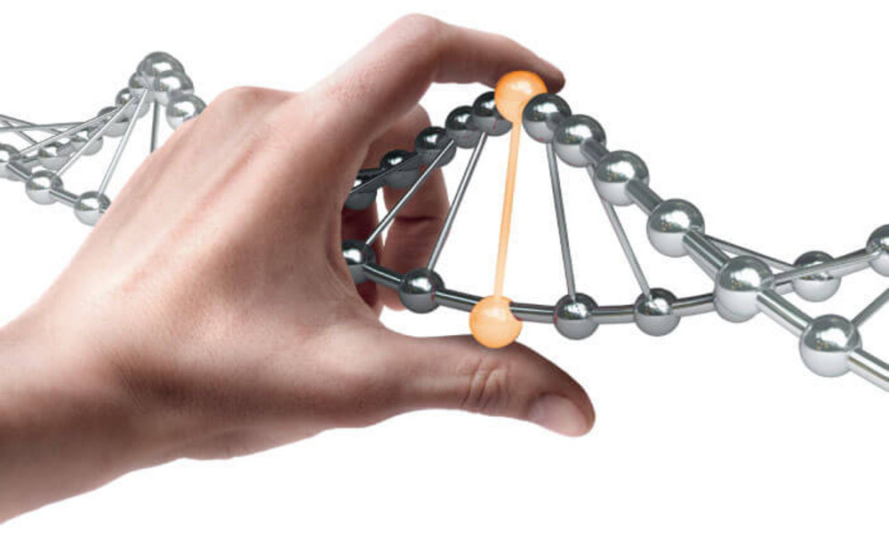
So, how does genetic testing work? First of all, a swab is used to collect DNA from the patient's mouth. The sample is then sent to Avellino Labs for next-generation sequencing and analysis (NGS). The custom NGS panel includes over 1,000 variants across 75 genes for keratoconus (KC) and over 70 TGFBI mutations for corneal dystrophies (CD). Sequencing results are aligned to Genome Reference Human Build 37 and a relative risk (RR) score is calculated for the detected keratoconus variant. Risk scores were derived from a Bayesian logistic regression model based on NGS results, including whole exome sequencing and targeted sequencing platforms.
The current treatments of cross-linking corneal are unable to repair the damage caused by keratoconus - changes to the cornea can cause deterioration of vision, this means that early diagnosis of keratoconus is a real need.

For surgery candidates, early diagnosis of keratoconus is very important as it can prevent post-operative progression of the disease. Holland explains: “By identifying those patients at risk earlier, we can improve monitoring for younger patients and potentially implement preventative treatments such as cross-linking. Genetic tests will also allow us to have further information in the evaluation of patients undergoing refractive surgery. Knowing a patient's potential to progress to keratoconus could be a deciding factor in choosing one refractive surgery procedure over another – or perhaps not recommending corneal refractive surgery."
csm-0719-700-webfirst-avellino-inarticle3-66af38fc9a_orig

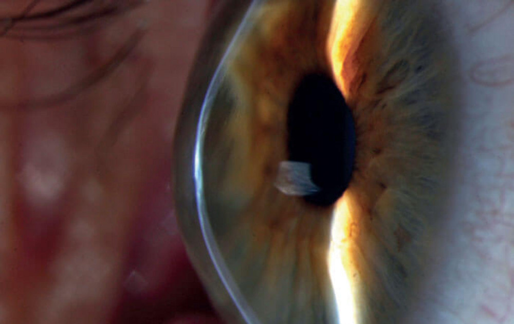
Eye with keratoconus, with visible central scarring and thinning with ectasia. Credits: David Yorston, Community Eye Health.

Corneal dystrophies, a contraindication for refractive surgery on a global scale

websitegraphic2-300dpi_1

Corneal dystrophies represent a strong contraindication to refractive surgery, unfortunately in some cases they are difficult if not impossible to diagnose: today there is an effective genetic test in 100% of cases.

Approximately 70 mutations related to the TGFBI gene causing corneal epithelial and corneal stromal dystrophies have been described to date, the genetic test Avellino detects the five most widespread types equal to 75% of the total.

A recent study collected data from 184 Human Gene Mutation Database (HGMD) and PubMed publications from 34 countries reporting 1600 cases of corneal dystrophies compared to 600,000 samples analyzed with genetic testing Avellino without finding any difference in mutation frequency in different populations or geographic locations, characterizing it as a global condition.(https://www.researchgate.net/publication/331070801_Evaluation_of_TGFBI_corneal_dystrophy_and_molecular_diagnostic_testing)

The genetic test Avellino detects the corneal dystrophies of Granular type I and II (or Avellino), Latex type I, Reis-Buckler and Thiel-Behnke, present in the world population with a prevalence of 1:500 to 1:1000, therefore a two-year pathology four times more common than keratoconus (https://www.sedesoi.com/vademecum3.php).

The medical literature has also demonstrated a strong correlation between PRK or LASIK interventions and a serious worsening of symptoms, even after years.

In subjects heterozygous for this mutation the signs are absent or difficult to diagnose, the test Avellino instead, it proved to be sensitive and specific to 100%: this means that a false positive or false negative was never detected in clinical trials.

For this reason the Italian Ophthalmological Society and the Italian Society of Legal Ophthalmology have expressed themselves in recommending the execution of the test both for medico-legal purposes and to allow the surgeon to evaluate alternative interventions, as well as representing the opportunity to offer safer refractive surgery to their patients.

Contact our product specialists for more information.

E-MAIL

Maculopathy, the 2RT laser proves effective in 3 out of 4 patients

og-image_2_orig

Age-related macular degeneration is the leading cause of blindness in industrialized countries, according to the LEAD study carried out by Center for Eye Research Australia (CERA) e published in Ophtalmology reveals for the first time an effective therapy.

Glaucoma? Finally free from eye drops with the SLT laser

shutterstock-eyedrops-1168x657-1_2

The prestigious journal The Lancet has published a three-year study on 718 patients by Prof. Gus Gazzard of Moorfields Eye Hospital which demonstrates how the SLT laser is more effective in treating glaucoma than eye drops, with possible enormous savings for healthcare public and a concrete improvement in the quality of life of patients.
Laser could soon replace eye drops in Britain to treat glaucoma patients, this is the surprising conclusion of a three-year study.

The results, published in the prestigious scientific journal The Lancet, show that Selective Laser Trabeculoplasty (SLT) laser technique should replace the use of eye drops as the main glaucoma therapy.

The study revealed that SLT is not only more effective and safer but could save the state coffers £1.5 million a year.

"A simple, safe, painless laser treatment that is not only more effective but also cheaper" says one of the authors, Prof. Gus Gazzard of Moorfields Eye Hospital from London.

"A single laser treatment relieves patients of the need to use eye drops every day for the rest of their lives, while also avoiding side effects."

Side effects associated with glaucoma drug therapies include cataracts, decreased heart rate, and fatigue.

Glaucoma is one of the main causes of blindness in the world, in Italy it affects a million people but half of them still don't know it and a late diagnosis seriously puts their sight at risk.

Most of these patients are prescribed eye drops to lower eye pressure that must be instilled every day for the rest of their lives, whereas SLT can be performed in 15 minutes and only once.

The patient sits in front of a microscope-like instrument and looks into it as 100 tiny pulses of light hit the eye, restoring the outflow of aqueous humor and lowering eye pressure.

The researchers organized a 3-year study of 718 patients and compared the effectiveness of the two treatments.

The results, published on the occasion of the World Glaucoma Week, clearly demonstrate that patients treated with SLT had lower and more stable eye pressure and fewer complications.

The use of SLT treatment could be decisive in countries without a developed healthcare system since it does not require the continuous use of drugs.

"This study represents a milestone," says Thomas Brunner, president of the Glaucoma Research Foundation.
“In developing countries, SLT could be a key tool for preventing blindness.”

en_USEnglish
Scroll to Top